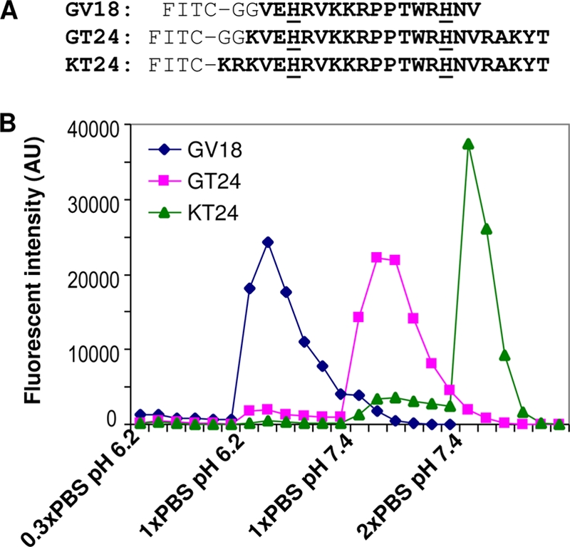
Fig 1.

Binding of peptides to heparin-agarose beads. (A) Sequences of the three peptides tested in this study for heparin binding. All peptides were labeled with FITC at the N termini. Residues from gp64 protein are shown in bold letters, and the two histidines are underlined. The two glycines serve as a spacer. GV18 contains only the disordered segment in the crystal structure of gp64. GT24 extends the sequence to the surrounding loop regions. In KT24, the two glycines in GT24 are replaced with two basic amino acids buried in the crystal structure of gp64. (B) FITC-labeled peptides were loaded onto a 1-ml heparin-agarose bead column and eluted with different elution buffers. Fractions of the elution were collected as 1 ml per fraction, and their fluorescence intensities were measured. AU, arbitrary units.
