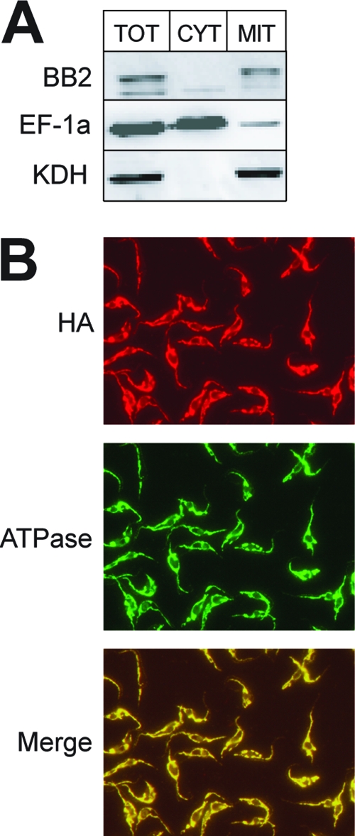
Fig 1.

Localization of epitope-tagged TbPPR9. (A) Immunoblot analysis of 0.3 × 10⁷ cell equivalents each of total cellular (TOT), crude cytosolic (CYT), and crude mitochondrial extracts (MIT) for the presence of the Ty1-tagged TbPPR9 protein (BB2). Only the relevant regions of the blots are shown. Comparison with molecular mass markers showed that the sizes of the tagged proteins were consistent with the prediction. eEF-1a served as a cytosolic marker (middle), and KDH was used as a mitochondrial marker (bottom). (B) Double immunofluorescence analysis of a T. brucei cell line expressing TbPPR9 carrying an HA tag. The cells were double stained with monoclonal anti-tag antibodies (HA) and a polyclonal antiserum directed against a subunit of the mitochondrial ATPase. A merged picture of the anti-tag antibody and the ATPase staining is also shown.
