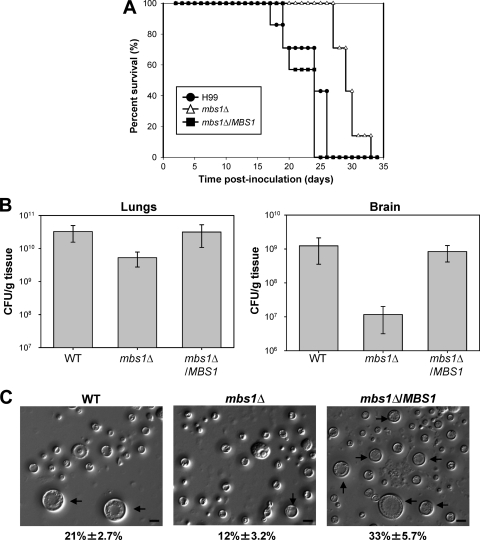
Fig 8.
Mbs1 is required for virulence of C. neoformans. (A) Groups of A/J mice were infected with 5 × 10⁴ cells of MATα WT (H99), mbs1Δ (YSB488), and mbs1Δ/MBS1 complemented (YSB1195) strains by intranasal inhalation. Survival was monitored for 33 days postinfection. (B) A/J mice were infected as indicated for panel A. Fungal burden (CFU/g tissue) was determined by plating homogenates of lung or brain tissue onto YPD at 21 days postinfection. P values for lungs: 0.05 (WT versus mbs1Δ), 0.949 (mbs1Δ/MBS1 versus WT), and 0.096 (mbs1Δ versus mbs1Δ/MBS1). P values for brains: 0.0747 (WT versus mbs1Δ), 0.5359 (WT versus mbs1Δ/MBS1), and 0.0302 (mbs1Δ/MBS1 versus mbs1Δ). (C) Mice were infected with 5 × 10⁶ cells of MATα WT (H99), mbs1Δ (YSB488), and mbs1Δ/MBS1 complemented (YSB1195) strains by intranasal inhalation. Cells were obtained by bronchial alveolar lavage at 3 days postinfection, fixed in formaldehyde, and analyzed by microscopy for size and morphology. More than 300 cells per animal were examined. Representative images are shown. Numbers indicate the percent titan cell formation ± SD. P values in pairwise comparisons: 0.0112 (WT versus mbs1Δ), 0.0294 (WT versus mbs1Δ/MBS1), and 0.0015 (mbs1Δ versus mbs1Δ/MBS1). Black arrows, titan cells. Bars = 10 μm.

