Figure 3.
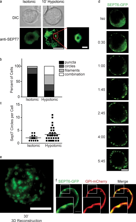
Septins assemble on the cell cortex during cortical retraction. (a) DIC and fluorescence images of wild-type D10 cells immobilized on anti-CD44–coated glass and stained for SEPT7 before or after a shift to 100 mOsm hypotonic media. Under isotonic conditions, SEPT7 is enriched at the cortex diffusely or in small puncta. Bar, 10 µm. (inset) Under hypotonic conditions, septins aggregate into filaments and rings. Bar, 1 µm. (b) Quantification of septin distributions observed in a. (c) Though some SEPT7 circles are observed in cells under isotonic conditions, each cell contains a greater number of rings under hypotonic conditions. Error bars represent SEM. (b and c) Representative data from one of two independent experiments performed. (d) Live imaging of SEPT6-GFP–expressing cells shifted to hypotonic media. Within 30 s, SEPT6-GFP begins to aggregate on the cortex and soon assembles into cortical rings that sometimes resemble tubules. Bar, 10 µm. (e) 3D reconstruction of SEPT6-GFP rings 20 minutes after a shift to hypotonic media. Bar, 10 µm. (f) SEPT6-GFP aggregations colocalize with invaginations in the plasma membrane, as indicated by GPI-mCherry fluorescence. Insets show boxed areas in greater detail. Bars, 2 µm.

