Figure 5.
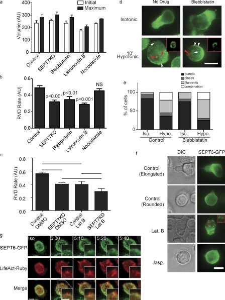
Interplay between septins and the actomyosin cytoskeleton in cell shrinkage. (a) Treatment with inhibitors of MyoII (blebbistatin), actin polymerization (latrunculin B), or microtubule polymerization (nocodazole) does not influence initial or maximum cell size in the flow cytometry osmotic swelling assay. AU, arbitrary unit. (b) Cortical retraction in this assay is slowed by blebbistatin or latrunculin but unaffected by nocodazole. (a and b) n = 7. (c) There is no additive or synergistic effect on cortical retraction of latrunculin B (Lat B) treatment of SEPT7KD cells. Lines indicate the groups between which statistical posttests were performed after analysis of variance. n = 5. (a–c) Error bars represent SD. (d and e) Illustration and quantification of anti-SEPT7 staining of wild-type cells showing that formation of septin filaments (white arrowheads) and rings (red arrowheads) is normal in cells treated with blebbistatin (pooled data from two independent experiments). Insets in d show septin rings in the boxed areas. Hypo, hypotonic; Iso, isotonic. (f) Imaging of SEPT6-GFP–expressing cells demonstrates septin aggregation into rings with latrunculin B treatment under isotonic conditions, whereas no such aggregation was observed with jasplakinolide (Jasp.) treatment. The inset shows latrunculin B–induced septin rings in greater detail. (g) Confocal imaging of cells expressing SEPT6-GFP and LifeAct-Ruby showing actin ruffles with subsequent recruitment of SEPT6-GFP to their bases. Bars, 10 µm.

