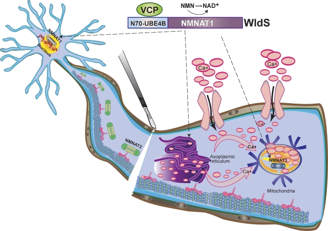
Figure 3.
A molecular model of WldS-mediated axon protection. The WldS fusion protein, consisting of the full-length Nmnat1 and the first 70 amino acids of Ube4b, is predominantly localized in the nucleus; however, it is also expressed in axonal cytoplasm and organelles such as mitochondria (broken arrows denote known neuronal sites of WldS expression) likely due to interaction with the cytoplasmic VCP protein. Expression of either WldS or extranuclear forms of Nmnat1 is sufficient to protect axons from degeneration upon injury, and this may result from substituting for the activity of Nmnat2 protein, which is degraded quickly after nerve injury. The WldS protein may also augment the enzymatic activity of Nmnat3, a mitochondrial Nmnat isoform, to confer axon protection. The combined result of ectopic Nmnat activities in WldS neurons may be less intracellular Ca2+ release from axoplasmic reticulum or greater Ca2+ buffering by the mitochondria via increased NAD+ production in these organelles, leading to overall decrease in intra-axonal Ca2+ levels (pink arrows denote net direction of Ca2+ flux).

