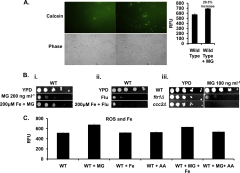
Fig 7.
Treatment with MG depletes LIP in C. albicans cells. (A) (Left) Measurement of LIP by using calcein in wild-type cells either left untreated or treated with MG (100 ng ml−1). (Right) Quantitation of fluorescence in wild-type cells either left untreated or treated with MG (100 ng ml−1). (B) (i) Serial dilution assays showing partial growth reversal upon supplementation with Fe (200 μM) in the presence of MG (200 ng ml−1). (ii) Serial dilution assays showing no growth reversal upon supplementation with Fe (200 μM) in the presence of FLC (2 μg ml−1). (iii) Serial dilution assay for the ftr1Δ and ccc2Δ iron transporter mutant strains in the presence of MG (100 ng ml−1). (C) Quantitation of fluorescence in the wild-type strain alone or in the presence of MG (100 ng ml−1), Fe (200 μM), AA (5 mM), MG plus Fe, or MG plus AA.

