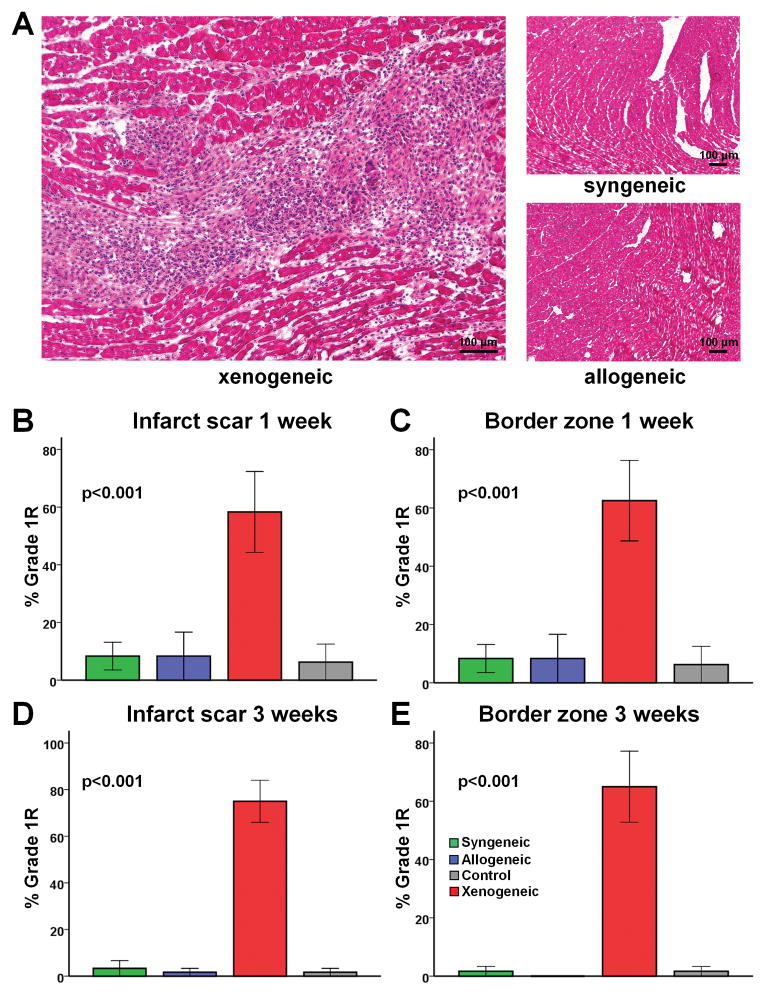
Figure 5.
Assessment of local immune rejection by H&E staining. (A) Representative images of H&E stained heart sections. No immune reaction can be detected in the allogeneic setting, while perivascular and interstitial mononuclear infiltration with no foci of myocyte damage can be observed in the xenogeneic setting (Grade 1R rejection). (B–E) Quantitative analysis of immune rejection based on the ISHLT grading system demonstrated that no significant immune rejection could be detected in the infarct scar and border zone 1 or 3 weeks after allogeneic cell transplantation. In contrast, xenogeneic cell transplantation resulted in Grade 1R rejection (n=4–5/group at each timepoint).

