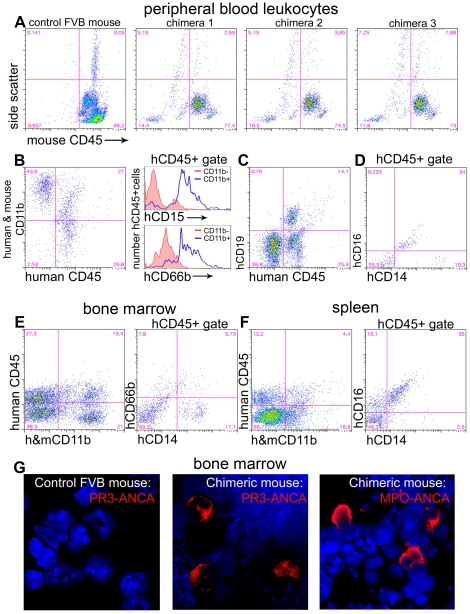
Figure 1. Characterization of chimerism in NOD-scid-IL2Rγ−/− mice.
(A–D) Flow cytometric analysis of leukocytes from tail bleeds six weeks after administration of HSCs (n = 26 mice). (A) Plots showing mouse leukocytes labelled with anti-mouse CD45 antibodies. Compared with control wild-type mouse blood, chimeras have populations of mCD45 negative leukocytes that show SSC characteristics of granulocytes (High), monocytes (Int) and lymphocytes (low). (B) Chimera blood leukocytes express human CD45 and many of these express CD11b. hCD45+,CD11b+ leukocytes predominantly express hCD15 and hCD66b compared with hCD45+,CD11b− leukocytes shown in histograms. (C) A proportion of hCD45+ leukocytes express CD19. (D) Some hCD45 leukocytes are CD14high and some are CD16+,CD14low. (E) In chimera bone marrow there are CD11b+ leukocytes which do not express mCD45 and among hCD45+ leukocytes a proportion express CD14 and a proportion express CD66b. (F) In chimera spleen there are CD11b+ leukocytes which express hCD45 and among hCD45+ leukocytes many express both CD14 and CD16. (G) Bone marrow spreads from wild type or chimera mice, labelled with anti-hMPO or anti-hPR3 IgG antibodies (red) purified from patients with vasculitis. Note that chimera bone marrow demonstrates anti-hMPO or anti-hPR3 antibody positive leukocytes with characteristic human neutrophil nuclear morphology. Wild type mouse bone marrow shows no cells positive for these antigens indicating that the anti-human antibodies do not cross react with mouse neutrophils.

