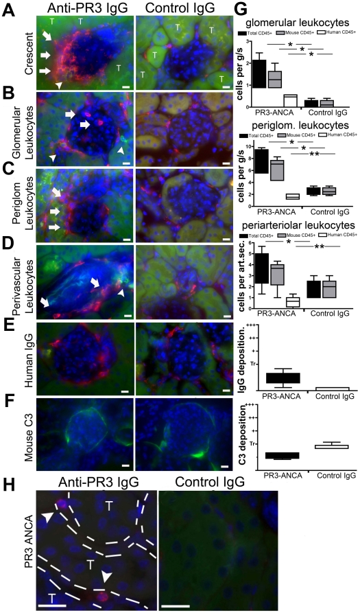
Figure 3. Anti-PR3 antibodies induce infiltration of kidneys with leukocytes of murine and human origin.
Kidney sections were incubated with anti-mCD45 (red) and anti-hCD45 (green) antibodies and images were captured by fluorescence microscopy (T = tubule). Occasional (<5%) glomeruli of anti-PR3 treated mice displayed intense extracapillary leukocyte infiltration (A) in the shape of crescents (arrows). Most glomeruli in animals treated with anti-PR3 antibodies (n = 18) had evidence of intraglomerular (B,G) and peri-glomerular (C,G) leukocyte infiltration. These were comprised mostly of mCD45+ cells, although some hCD45 leukocytes were also present (arrowheads). In addition, there was a significant increase in peri-vascular leukocyte (mCD45+ and hCD45+) infiltration in anti-PR3 treated mice (D,G [per arteriolar section (art.sec.)]). Sections were also stained for deposition of IgG [red] (E,G) and C3 [green] (F,G). IgG was detectable within periglomerular cells, but there was minimal deposition within the glomeruli. Mouse C3 was weakly deposited in glomeruli but was no different between control group (n = 8) and anti-PR3 group (n = 18). Note mouse C3 can be detected normally binding avidly to tubular basement membranes. (Marker = 10 µm) (*P<0.05, **P<0.01. median ± IQ ± max/min values). (H) Kidney sections from anti-PR3 and control treated animals were incubated with anti-PR3 positive ANCA IgG. In the peritubular capillaries of chimera mice that received anti-PR3 hIgG occasional leukocytes detected by anti-hPR3 hIgG could be detected. No positively stained human neutrophils were seen in glomeruli.

