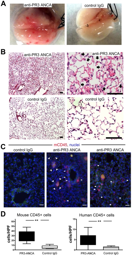
Figure 4. Anti-PR3 antibodies induce capillaritis with leukocytes of mouse and human origin.
(A) Photomicrographs of explanted lungs from mice treated with disease control IgG (n = 8) or from patients with anti-PR3 ANCA (n = 18). Mice treated with anti-PR3 ANCA displayed petechiae over the surface of the lung. (B) H&E stained low and high power images of lungs from chimera mice injected with anti-PR3 antibodies 6 days previously. Note hemorrhage, inflammation, thickening of alveolar walls, enlarged, highly vacuolated alveolar macrophages and prominent recruitment of neutrophils (arrows) within the alveolar walls. These features are consistent with capillaritis. By comparison chimera mice treated with control IgG showed normal lung architecture. (C) Immunostaining of lung tissue to assess the degree of leukocyte infiltration. Note that in mice that received anti-PR3 ANCA IgG there was significant recruitment of leukocytes to the peribronchial areas (thin arrows) and also the alveolar areas (fat arrows). In addition many alveoli in mice treated with anti-PR3 antibodies had apoptotic debris that was partially ingested by alveolar macrophages (arrowheads) (bar = 50 µm). (D) Blinded assessment of human and mouse leukocyte recruitment to the lungs of treated animals (** P<0.01. median ± IQ ± max/min values).

