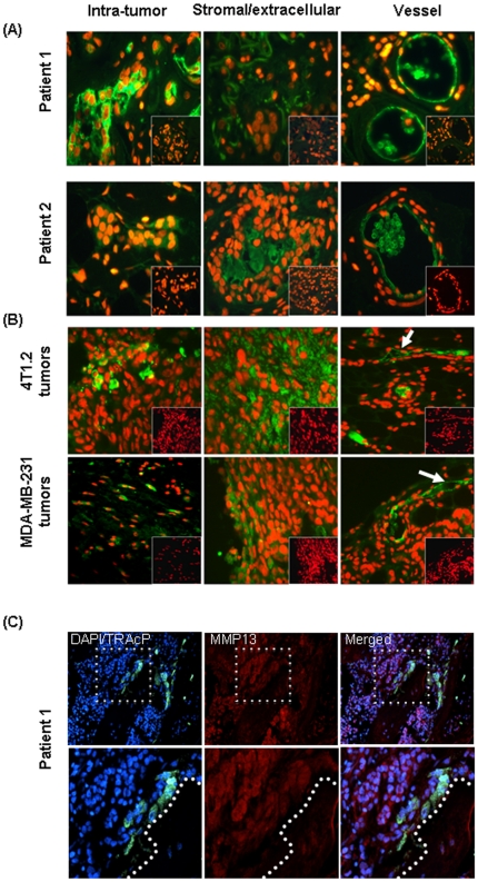
Figure 8. Immunofluorece for MMP13.
(A) MMP13 immunofluorescence images of the clinical breast tumors from Patients 1 and 2. Immunofluorescence staining was performed on FFPE sections for MMP13 using a sheep polyclonal MMP-13 antibody as indicated by green fluorescence, whereas nuclei are stained red with propidium iodide. Parallel sections from the same tissue blocks were probed with control sheep serum to monitor for non-immune background (lack of green signal displayed as an inset in each panel). (B) Representative images of MMP13 immunofluorescence (green) from the syngeneic (top panel) and xenograft (bottom panel) mouse tumors. (C) MMP13 immunofluorescence (red) in human breast to bone metastasis. Dashed box in upper panels represents area of magnification in lower panels. Multinucleated (DAPI; blue) osteoclasts (TRAcP; green) were identified at the tumor-bone interface (represented by dashed line in the lower panels).

