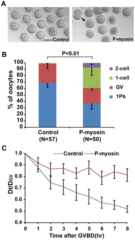
Figure 7. Microinjection of oocytes with monoclonal anti-phospho-myosin antibody facilitated abnormal cytokinesis in meiosis I.
(A) Representative images of oocytes taken 16 hours after microinjection with DAM-488 2nd antibody (Control) and anti-phospho-MLC2 (Ser19) antibody (P-myosin). The arrowhead indicates a symmetrically dividing oocyte. Scale bar: 100 µm. (B) The frequency of 1 Pb, GV, 1-cell and 2-cell type divisions in the Control and P-myosin groups. The P value was calculated using a 2×4 χ2-test. (C) The shortest distance between chromosomes and the cortex was measured in 15 oocytes per group at GVBD (DGV) and hourly time points after GVBD (Dt). Dt/DGV plotted against time was used as an indicator of chromosome movement.

