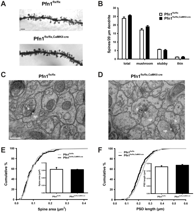
Figure 2. Unaltered spine density and morphology in hippocampal CA1 region of Pfn1flx/flx,CaMKII-cre mice.
(A) Representative images of 2nd order dendritic branches of Golgi-stained pyramidal cells in the hippocampal CA1 stratum radiatum. Scale bar: 2 µm. (B) Unaltered spine density in Pfn1flx/flx,CaMKII-cre mice. Spines were morphologically categorized into mushroom-like, stubby, and thin spines (>1,000 µm length of dendritic branches for both groups, four mice per group). Representative electron micrographs of CA1 stratum radiatum of (C) Pfn1flx/flx controls and (D) Pfn1flx/flx,CaMKII-cre mice. Scale bar in C: 200 nm. b: presynaptic bouton, sp: dendritic spine, *: postsynaptic density. Unaltered spine area (E) and PSD length (F) in Pfn1flx/flx,CaMKII-cre mice as deduced from cumulative distributions and mean values (insets in E and F).

