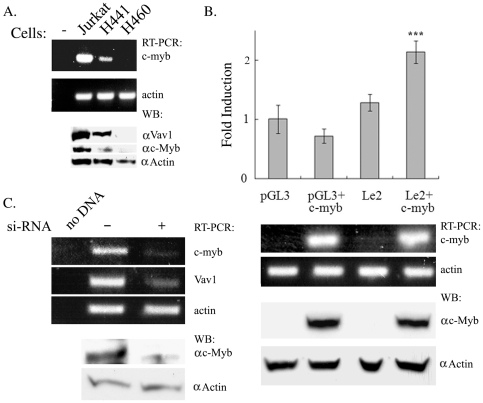
Figure 5. C-Myb is involved in regulation of vav1 expression in lung cancer cells.
(A) Endogenous expression of c-myb mRNA in Jurkat T cells, H441 (vav1-positive) and H460 (vav1-negative) lung cancer cell lines was detected by RT-PCR and western blotting. (B) Empty vector pGL3 or the Le2 wt reporter construct was transfected either alone or with a c-Myb-expressing plasmid into H460 lung cancer cells (as in Materials and Methods). Luciferase activity was measured 24 hr after transfection (top panel). Luciferase activity is expressed as fold induction relative to basic pGL3 expression. Values are the mean of five independent experiments; significance was determined using the unpaired student T test. (***) indicates p<0.01. The bottom panel shows the level of c-myb and actin mRNA and protein expression in the transfected cells as determined by RT-PCR and Western blotting respectively. (C) H441 lung cancer cells were transfected with either scrambled DNA (-) or with siRNA against c-Myb. Seventy-two hours later, the mRNA levels of c-myb, vav1 and actin were detected by RT-PCR.

