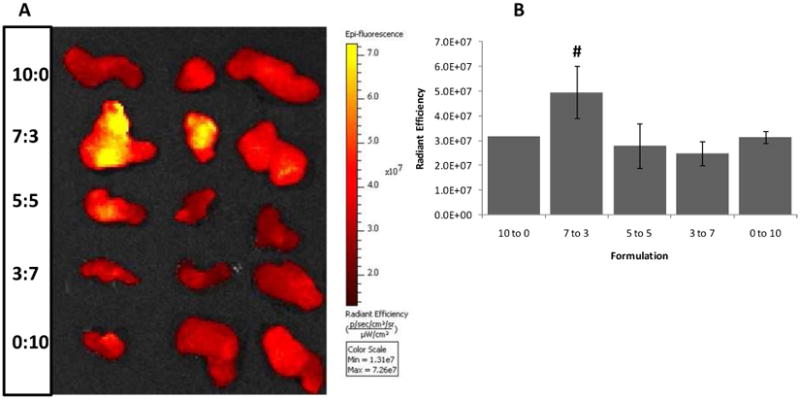
Figure 7.

Excised tumor tissues following sacrifice of mice injected with ICG-encapsulating mixed micelles at various PEO-PHB-PEO to PF127 ratios. (A) Fluorescence image of excised tumor tissues obtained 24 hr after initial injection of ICG. (B) Quantification of ICG fluorescence in tumor tissue excised 24 hr after injection using ROI analysis. # indicates a statistically significant difference between the 7:3 mixed micelles and 10:0 micelles (one-tailed p=0.035, n = 3).
