
Figure 4.
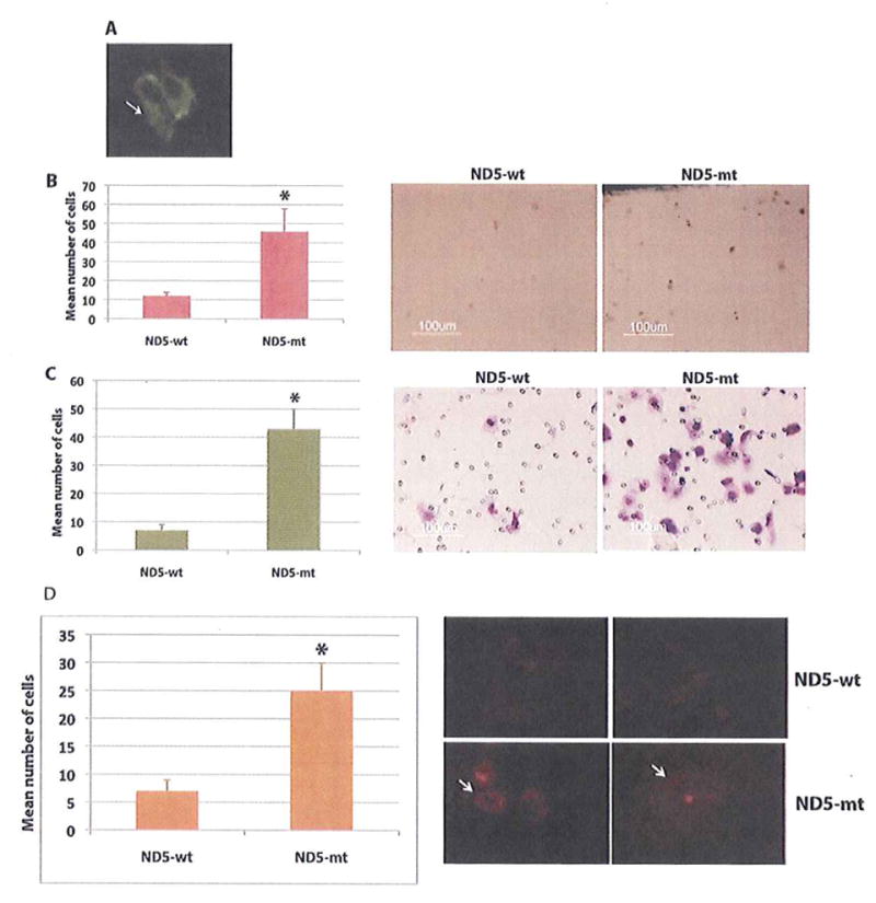
Impact of forced overexpression of ND5 gene on in vitro proliferation, invasion and superoxide production of lung cancer cells SW-900. A. The presence of the exogenous fusion protein in the mitochondria of the ND5 transfected cells was detected using anti-myc antibody by immunofluorescence (arrow). B. Number of proliferating cells was significantly higher (P=0.0031, indicated by asterisk) in the ND5 mutant (ND5-mt) transfected cells compared to the wild type (ND5-wt) transfected cells (Left panel). Representative photomicrograph from each experimental group is shown in the right panel. Magnification X 200. C. Number of invasive cells was also significantly higher (P=0.0002, indicated by asterisk) in the ND5 mutant (ND5-mt) transfected cells compared to the wild type (ND5-wt) transfected cells (Left panel). Representative photomicrograph from each experimentai group is shown in the right panel. Magnification X 400 C. D. Number of superoxide producing cells was determined by the MitoSox superoxide assay. Number of positive cells was significantly higher (P=0.0049, indicated by arrow) in the ND5 mutant (ND5-mt) transfected group compared to the wild type (ND5-wt) transfected group (Left panel). Representative photomicrograph from each experimental group is shown in the right panel. Magnification X 200.