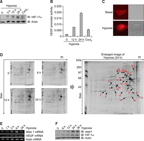
Figure 1.
Hypoxia induced Wee1 expression. (A) Induction of HIF-1α by hypoxia in MS-1 cells. Cells were exposed for the indicated times in a hypoxic chamber (0.1% O2) and then blotted with HIF-1α antibody. The lower panel demonstrates equivalent loading of total actin in the whole cell lysates. (B) The effect of hypoxia on the VEGF promoter. The promoter plasmid was transiently transfected and maintained for 24 h in MS-1 cells. After hypoxia (0.1% O2) for indicated times, the promoter activity was monitored. CoCl2 was treated and used as a chemical hypoxic inducer. (C) Effect of hypoxia on subcellular localization of HIF-1α. Before hypoxic treatment, MS-1 cells were preincubated for 30 min with hypoxia culture medium. The HIF-1α signal was then measured after staining with antibody. (D) Spectrometric analysis of hypoxic samples. Two-dimensional gels showing the silver-stained signal obtained from 200 µg of MS-1 cell before and at measured intervals after induction of hypoxia. The right image is the enlarged images of the 24 h spot in the 2D gels. The arrows indicate the identification of the protein by spot-picking, tryptic digestion, and MALDI-TOF. (E) Image of total RNA was prepared from MS-1 cells and amplified by RT-PCR using Wee1/VEGF-specific primers. Actin mRNA was used as a positive control. The PCR products were separated on 2% agarose gels and visualized with UV. (F) Effect of hypoxia on Wee1 expression. The MS-1 cells were exposed to hypoxia for the indicated times. Expression of Wee1 was then examined by blotting with antibodies to HIF-1α and Wee1. The lower panel demonstrates equivalent loading of total actin in the whole cell lysates.

