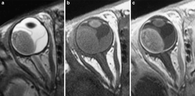
Fig. 3.
Transaxial T2-weighted (TR/TE, 3,460/116 ms) (a) and T1-weighted (TR/TE, 374/14 ms) precontrast (b) and postcontrast (c) MRI of exophytically growing retinoblastoma with secondary retinal detachment. Retinoblastoma typically has low signal intensity compared to the vitreous body on T2-weighted images and intermediate signal intensity on precontrast T1-weighted images, and it demonstrates marked contrast enhancement

