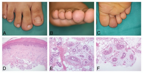
Figure 1.
Clinical appearance of the skin lesion. (A–C) Swelling of the plantar surface was apparent on the right second toe. The overlying surface was erythematous with a small amount of fine scales. Histologically, the epidermis showed acanthosis with hypertrophy of the granular layer and mild elongation of rete ridges. (D) A mild, perivascular inflammatory infiltration in the upper dermis was seen (hematoxylin and eosin, original magnification x40). (E) A nodular proliferation of normally structured eccrine coils and ducts in the deep dermis and subcutaneous fat tissue. Some of the ducts show mild dilation. Extravasation of erythrocytes was also noted around the proliferation of eccrine glands (hematoxylin and eosin, original magnification x200). (F) Proliferation of small capillary-sized vessels in the stroma surrounding the eccrine glands in the subcutaneous fat tissues (hematoxylin and eosin, original magnification x200).

