Figure 1.
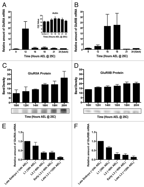
Expression of Drosophila GluRIIA and GluRIIB mRNA and protein through embryonic and larval development. (A) Amount of GluRIIA mRNA in wildtype (Oregon R) embryos, measured using quantitative real time RT-PC R. Quantity is presented relative to the amount measured at 24 h after egg laying (AEL). N = 5–10 independent mRNA isolations and measurements per time point. Actin 5C mRNA was amplified and measured concurrently with GluRs as a control for mRNA isolation and amplification. Inset: Actin 5C raw C(t) values from unfertilized (unfert.) eggs laid by virgin females, and at various time points during embryogenesis. Note the lack of significant variation. (B) Amount of GluRIIB mRNA during embryogenesis, measured and quantified as described for GluRIIA. (C) Amount of GluRIIA protein, as measured by quantitative immunoblots. N = 3–8 independent protein isolations and measurements per time point. (D) Amount of GluRIIB protein, as measured by quantitative immunoblots. N = 3–8 independent protein isolations and measurements per time point. (E) Amount of GluRIIA mRNA in hatch-age (24 h AEL) embryos, first instar (L1), second instar (L2) and third instar (L3) larvae. N = 3 independent mRNA isolations and measurements per time point. (F) Amount of GluRIIB mRNA in hatch-age embryos and larvae, as described for (E). N = 3 independent mRNA isolations and measurements per time point.

