Figure 4.
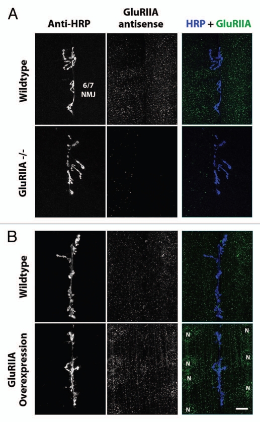
GluRIIA mRNA aggregates in third instar larval muscles. (A) Top parts: Confocal micrographs of ventral longitudinal muscles 6 and 7 in one hemisegment of a wild type third instar larvae. Left column shows NMJs, visualized using fluorescently-conjugated anti-HRP. Middle column shows anti-GluRIIA FISH signal. Right column shows merge of anti-HRP (blue) and anti-GluRIIA mRNA (green) signal. Bottom parts: As top, but showing hemisegments from a GluRIIA[AD9]/Df(2L) Exel8016 mutant larva, in which the GluRIIA gene is deleted. (B) Top parts: Confocal micrographs of third instar ventral muscles 6 and 7, as in (A). Bottom parts: As top, but showing hemisegments from a [UAS-GluRIIA/+; 24BGal4/+] mutant larva, in which GluRIIA is overexpressed specifically in muscle cells. Muscle nuclei are labeled with letter ‘N’. Scale bar: 15 um.

