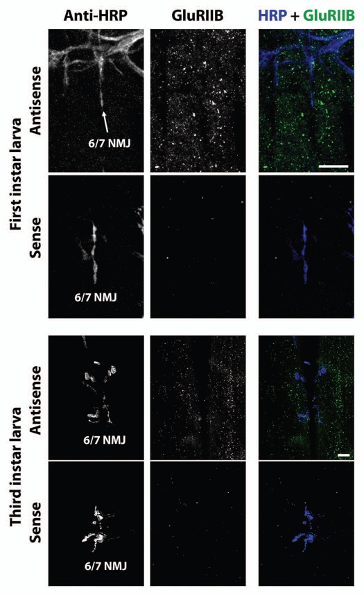
Figure 6.
GluRIIB mRNA aggregates in first and third instar larval muscles. Top six parts: Confocal micrographs of first instar NMJs and muscles as in Figure 3, except using antisense probes against GluRIIB instead of GluRIIA. Bottom parts show (lack of) FISH signal resulting from use of sense (negative control) probes. Lower six parts: Confocal micrographs of third instar NMJs and muscles as in Figure 4, except using antisense probes against GluRIIB instead of GluRIIA. Bottom parts show (lack of) FISH signal resulting from use of sense (negative control) probes. Scale bars: 15 um.

