Figure 8.
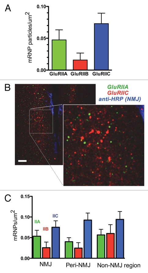
Different GluR mRNAs are associated with different mRNA aggregates. (A) mRNA aggregate density in third instar larval muscles 6 and 7, for GluRIIA, GluRIIB and GluRIIC. Values are ‘background-subtracted’ such that the number of punctae in sense controls is zero. (B) Confocal micrographs of third instar ventral longitudinal muscles 6 and 7, after multiplex FISH and simultaneous immunohistochemistry to visualize the NMJ. GluRIIA mRNA aggregates are green; GluRIIC mRNA aggregates are red, and the NMJ is blue. Note that the GluRIIA and GluRIIC mRNA aggregates vary in density and do not overlap. Scale bar: 15 um. (C) GluRIIA, GluRIIB and GluRIIC mRNA aggregate density in muscles 6 and 7, separated according to distance from the NMJ. ‘NMJ’ density was measured in the area delimited by anti-HRP staining. ‘Peri-NMJ’ density was measured outside the anti-HRP staining but within 10 um of the NMJ. ‘Non-NMJ region’ density was measured in muscle areas greater than 10 um from the NMJ. N = 9–14 animals per measurement.

