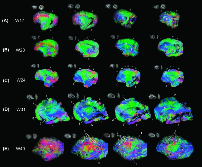
Figure 2.
Tractography pathways at W17 (A), W20 (B), W24 (C), W31 (D), and W40 (E). (A) Tractography at W17—dominant radial pathways with immature forms of projection, limbic, and few emergent association pathways. a: radial pathways (in dorsal areas), b: radial pathways (in ventral areas), c: radial pathways (medio-lateral horizontal direction), d: corticospinal tract, e: corticothalamic/thalamocortical tract, f: fornix, g: ganglionic eminence, j: uncinate fasciculus, k: inferior fronto-occipital fasciculus. We put the label (k) deep inside from the temporal lobe where the tract is running, which in the sagittal view looks running through the temporal lobe, but the tract is actually running between the frontal and occipital cortices. (B) Tractography at W20—dominant radial pathways and emerging long-range connectivity patterns. a: radial pathways (in dorsal areas), b: radial pathways (in ventral areas), c: radial pathways (medio-lateral horizontal direction), e: corticothalamic/thalamocortical tract, f: fornix, g: ganglionic eminence, j: uncinate fasciculus, k: inferior fronto-occipital fasciculus, m: superficial horizontal pathways, n: frontotemporal pathways. Similar features were noted in W22 specimen (not shown). (C) Tractography at W24—less predominant radial pathways in dorsal frontal, parietal and inferior frontal lobes, and emergent short-range corticocortical and long-range association pathways. a: radial pathways (in dorsal areas), b: radial pathways (in ventral areas), e: corticothalamic/thalamocortical tract, f: fornix, g: ganglionic eminence, j: uncinate fasciculus, o: corticocortical pathways (in parietal and parieto-fontal areas), p: corticocortical pathways (in parietal areas without gyrus), q: corticocortical pathways (in ventral frontal areas). (D) Tractography at W31—less dominant radial pathways in the temporal and occipital lobes, emergent short-range corticocortical and long-range association pathways ventrally. a: radial pathways (in dorsal areas), b: radial pathways (in ventral areas), e: corticothalamic/thalamocortical tract, f: fornix, g: ganglionic eminence, h: cingulum, j: uncinate fasciculus, k: inferior fronto-occipital fasciculus, p: corticocortical pathways (in parietal areas without gyrus), q: corticocortical pathways (in ventral frontal areas), r: corticocortical pathways (in superior frontal areas), s: superior fronto-occipital fasciculus, t: cortical u-fibers, u: radial pathways between gyral apices and ventricular regions. (E) Tractography at W40—no evident radial pathways, abundant complex, crossing short- and long-range corticocortical association pathways. e: corticothalamic/thalamocortical tract, f: fornix, k: inferior fronto-occipital fasciculus, t: cortical u-fibers, v: corticocortical pathways (between the insula and temporal cortex), w: inferior longitudinal fasciculus. Similar features were noted in W38 specimen (not shown). In all panels, we show tractography pathways that pass through a sagittal slice that is shown as an insert in upper left corner. In a coronal slice next to the sagittal slice, the location of the sagittal slice is shown as a yellow line. For visualization purposes in the figures, we restricted the percentages of the number of detected tractography pathways in the following ways. We showed only 70% of the tractography fibers that touched each sagittal slice and were more than 2 mm in length. The label g actually points at tangential pathways in the ganglionic eminence structure running in the anterior–posterior direction (shown in blue). Colors indicate spatial relationships between fiber end points. Red: left–right, blue: anterior–posterior, and green: dorsal–ventral orientation. As we explain in the text, callosal connections in the specimens were disrupted at the midline at autopsy, so callosal tractography pathways appeared in green (dorsoventral direction: from the corpus callosum to the cortex).

