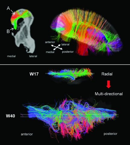
Figure 5.
Upper panel: Tractography pathways at W22. Colors indicate spatial relationships between fiber end points. Red: left–right, blue: anterior–posterior, and green: dorsal–ventral orientation. As we explain in greater detail in the text, callosal connections in the specimens were disconnected at the midline during craniotomy, so callosal tractography pathways appeared in green (dorsoventral direction: from the corpus callosum to the cortex). (A) Corticocortical pathways (in dorsal areas including occipital, parietal, and frontal areas). (B) Callosal pathways. We selected only 2 types of pathways of interest using appropriate slice filters. Lower panel: axial views of fiber pathways at W17 and at W40. Only fibers that pass a sagittal plane (shown as a white slice in the Figure) are shown.

