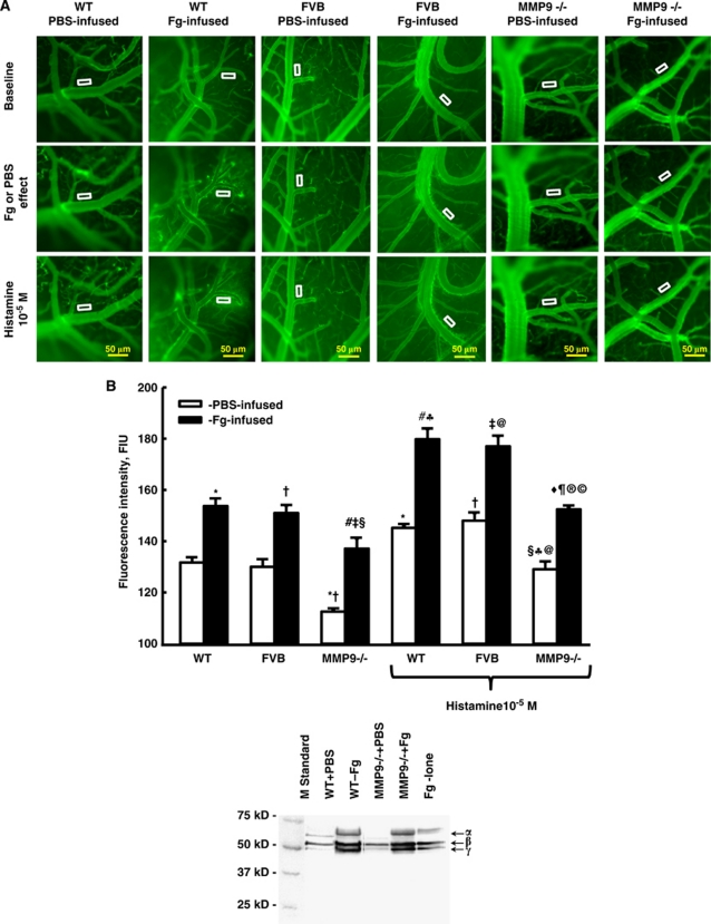
Figure 2.
Fibrinogen (Fg)-induced macromolecular leakage of pial venules. (A) Examples of images recorded before (baseline; first row), after infusion of Fg (final blood concentration 4 mg/mL) or phosphate-buffered saline (Fg or PBS; second row), and topical application of histamine (10−5 mol/L) (third row), in wild-type (WT; first two columns), FVB (second pair of columns), and matrix metalloproteinase-9 (MMP-9) gene knockout (MMP9−/− third pair of columns) mice. Microvascular leakage was assessed by fluorescence intensity of fluorescein isothiocyanate-bovine serum albumin (FITC-BSA) in the rectangular area of interest (AOI) shown on images. (B) Summary of changes in fluorescence intensity after infusion of Fg or PBS measured in the AOI. P<0.05 for all. *—versus WT-PBS, #—versus WT-Fg, †—versus FVB-PBS, ‡—versus FVB-Fg, §—versus (MMP9−/−)-PBS ⧫—versus (MMP9−/−)-Fg, ♣—versus WT+PBS/Hist, ¶—versus WTFg/Hist; @—versus FVB-PBS/Hist, ®—versus FVB-Fg/Hist, ©—versus MMP-PBS/Hist. n=8 for all groups. Inset: Purity of Fg with no visible extra bands lighter than ∼49 kDa (that would indicate degradation products) was confirmed by the Coomassie-stained sodium dodecyl sulfate polyacrylamide gel electrophoresis (SDS-PAGE) analysis.

