Figure 5.
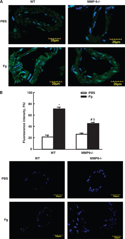
Activation of matrix metalloproteinases (MMPs) in mouse pial vessels. (A) Examples of vessel images in samples obtained from wild-type (WT; first column) and MMP-9 gene knockout (MMP9−/− second column) mice infused with phosphate-buffered saline (PBS; first row) or fibrinogen (Fg; second row). MMP activity was assessed by fluorescence intensity (green) along the pial vascular segment. 4,6-Diamidino-2-phenyl-indole (DAPI)-labeled cellular nuclei are shown in blue. (B) Summary of fluorescence intensity changes in the brain vessels after infusion of Fg or PBS. P<0.05 for all. *—versus WT+PBS, #—versus WT+Fg, †—versus (MMP9−/−)+PBS; n=4 for all groups. Inset: Validity of the test was confirmed in parallel series of experiments done on WT and MMP9−/− mice. The brain cryopreserved slices were treated with 1,10-phenanthroline,monohydrate a general metalloproteinase inhibitor (green) as a negative control. Cell nuclei are labeled with DAPI (blue). The color reproduction of this figure is available on the Journal of Cerebral Blood Flow and Metabolism journal online.

