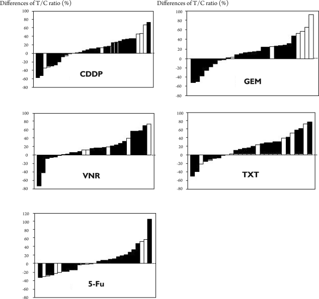
Figure 2. Differences in the T/C ratio (%) between the primary lesions and their paired metastatic lesions.
Differences in the T/C ratio (%) between the primary lesions and their paired metastatic lesions are shown by waterfall plots. The black columns show the difference between primary lesions and their paired lymph node lesion. The white columns show the difference between primary lesions and their paired non-lymphatic lesions. Although the cases involving a non-lymphatic route (white column) were widely distributed, 4 cases (67%) treated with GEM showed a 30% or greater increase in in vitro resistance in the paired metastatic lesions compared with the primary tissue. Three cases (50%) showed similar results for CDDP.

