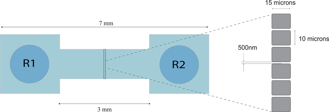
Figure 2.
Schematic of a nanofluidic flow-cell fabricated on fused silica wafers, showing reservoirs R1 and R2 at its ends. Virus sample is introduced into reservoir R1 and are made to flow through the flow-cell by pressure-driven flow. Nanochannels are present in the zoomed in area of the flow-cell. Nanochannels (cross section 500 nm × 400 nm, length 15 µm) form an array across the 15 µm ridge along the center of the flow-cell, as shown in the figure. The laser focus is placed at the center a nanochannel to detect the passage of individual viruses or nanoparticles.

