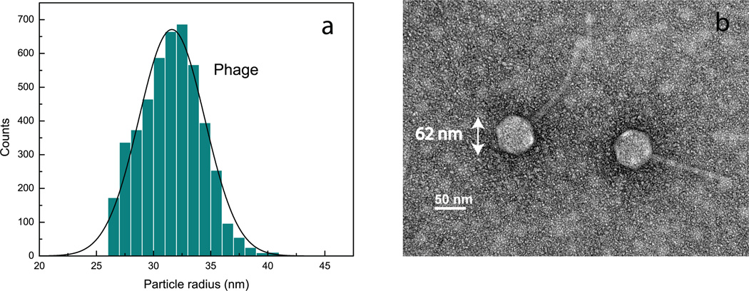
Figure 6.
Analysis of a bacteriophage lambda sample.(a) Size distribution obtained using dark-field interferometric detection.(b)TEM micrograph from the sample used in (a). The hexagonal head along with the tail makes a phage particle easily recognizable in the image. Radii of the heads of the phage particles (half the distances between opposite corners of the hexagons) are determined from the image to be 31 nm, in close agreement with the mean size determined from our optical measurements.

