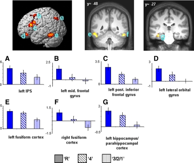
Figure 2.
(Upper) Modality-independent subsequent memory effects (main effect of memory [P < 0.001] exclusively masked with subsequent memory by modality interaction [P < 0.1]). Effects are rendered onto a single subject template brain (left) and projected onto a section of the normalized average anatomical image (right). (Lower) Bar plots show parameter estimates (in arbitrary units) for recollected (R), familiar (4), and forgotten (3/2/1) trials of peak voxels for effects localized in (A) left IPS (−30, −69, 42), (E,F) left (−51, −48, −21) and right fusiform cortex (51, −48, −18), (B) left middle frontal gyrus (−27, 9, 51), (C) left posterior inferior frontal gyrus (−45, 18, 15), (D) left lateral orbital gyrus (−39, 39, −18), and (G) left hippocampus/parahippocampal cortex (−39, −27, −18).

