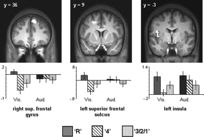
Figure 4.
(Upper) Visually selective subsequent recollection effects projected onto sections of the normalized across-subjects averaged anatomical image. (Lower) Bar plots of mean parameter estimates (in arbitrary units) for recollected (R), familiar (4), and forgotten (3/2/1) trials in right superior frontal gyrus, left superior frontal sulcus, and left insula.

