
Figure 3.
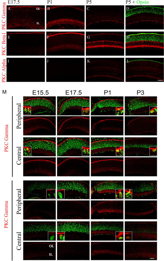
PKC-β1 and PKC-γ isoforms are expressed during retinal development. A–L, Immunofluorescent detection of PKC-α, PKC-β1, and PKC-γ (red) from E17.5 (A, E, I), P1 (B, F, J), P5 (C, G, K) and colocalization with opsin (green) expression at P5 (D, K, L). M, Expression of PKC-β1 and PKC-γ (red) colocalization with PCNA (green) in the central and peripheral retina of E15.5, E17.5, P1, and P3 retinas. OL, Outer retinal layer; IL, inner retinal layer. Scale bar, 40 μm.