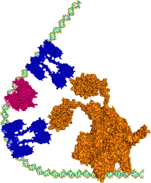
Fig 5.
Schematic representation of the bending of phhA promoter induced by the specific binding of two PhhR dimers and Crp protein. The PhhR dimers are colored in blue and Vfr is in magenta; RNA polymerase is in orange. The PhhR proteins are bound to the distal and the proximal phhR binding boxes, and Crp is bound to its binding site, allowing the PhhR dimers to be in close proximity to RNA polymerase.

