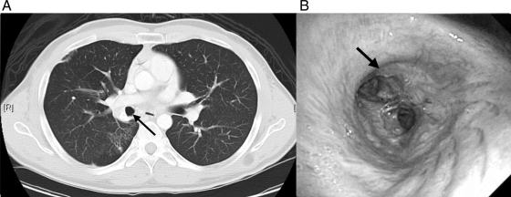
Fig 3.
(A) Chest CT 10 months after presentation. Interval clearing of dense RLL consolidation and the decreased size of the partially calcified mass in the right hilum can be seen. The patent bronchus intermedius (arrow) was viewed by CT (A) and follow-up bronchoscopy (B) after laser ablation and mechanical debulking.

