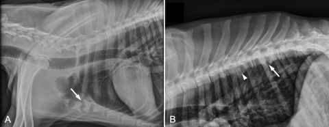
Fig 1.
(A) Lateral radiograph of the thorax. There is lysis of the first four sternebrae and marked shortening of the second and third sternebrae, which have irregular margins and loss of the end plates (arrow). (B) Lateral radiograph of the thoracic vertebral column. There is end plate lysis of the 9th (T9) and 10th (T10) thoracic vertebrae that is centered on the intervertebral space (arrow), with spondylosis deformans ventrally. There is also narrowing, end plate sclerosis, and spondylosis deformans between the seventh (T7) and eighth (T8) vertebrae (arrowhead).

