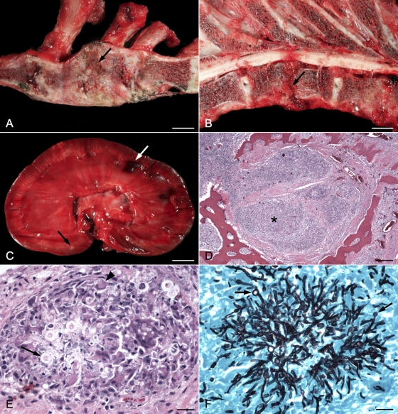
Fig 2.
(A) Right sagittal section of sternum; the first sternebra is on the left. The second and third sternebrae are collapsed, and areas of bony proliferation obscure the joint space. An area of necrotizing osteomyelitis partially separates the two sternebrae (arrow). Bar, 1 cm. (B) Left sagittal section of thoracic vertebrae; the cranial end is to the right. The intervertebral disk at T9-T10 is missing (arrow). The end plates are eroded, and a wedge-shaped piece of tissue compresses the spinal cord dorsally. Bar, 1 cm. (C) Sagittal section of left kidney. Small white areas are scattered throughout the cortex and medulla (black arrow). The pelvis is dilated, and the renal crest is ulcerated. Areas of hemorrhage (white arrow) are visible in the cortex. Bar, 1 cm. (D) Photomicrograph of second sternebra showing areas of inflammation (*) and surrounding fibrous tissue invading and replacing the marrow cavity. Bar, 250 μm. (E) Higher magnification of sternebra showing marked granulomatous inflammation with giant cell formation (arrowhead) surrounding septate fungal hyphae and bulbous spore-like structures (arrow). Bar, 25 μm. (F) Grocott's methenamine silver-stained section of sternebra taken from same area as previous image, demonstrating prominent fungal hyphae and terminal conidiophores (arrows). Bar, 25 μm.

