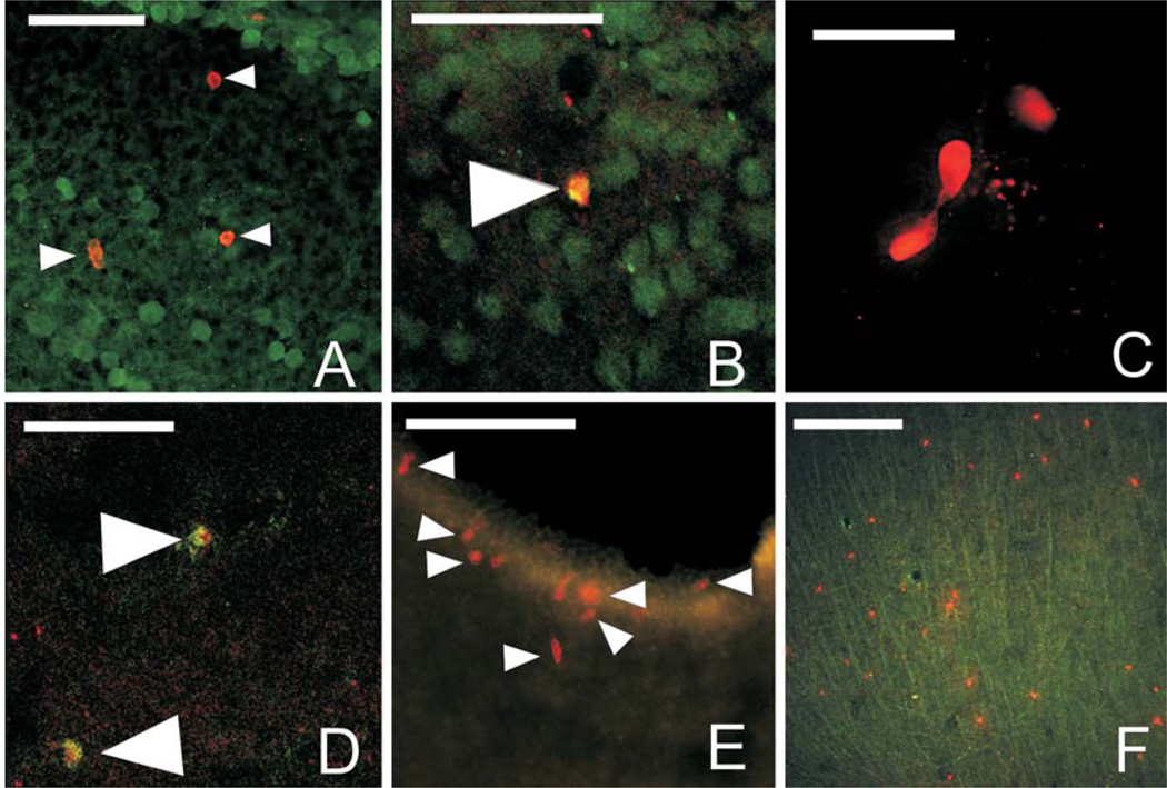
Fig. 3.
BrdU label and double label in various regions of the adult brain. A Discrete label of BrdU- (red, solid white arrowheads) and GFAP- (green) labeled cells in the laminar nucleus of the TS 7 days post-BrdU injection. B Confocal image of BrdU/GFAP-double-labeled cell (solid white arrow) in the ventral hypothalamus 7 days post-BrdU injection. C Dividing BrdU-labeled cells (red) in the posterior nucleus of the thalamus at 14 days post-BrdU injection. D Confocal image of BrdU/TOAD-64-double-labeled cells (solid white arrowheads) in the posterior nucleus of the thalamus 14 days post-BrdU injection. E BrdU-labeled cells (red) around the ventral portion of the telencephalic ventricle (region of the nucleus accumbens) 21 days post-BrdU injection. F BrdU-labeled cells (red) and GFAP-labeled fibers (green) in the optic tectum 28 days post-BrdU injection. For all images, medial is to the left and dorsal is up. Scale bars = 50 µm.

