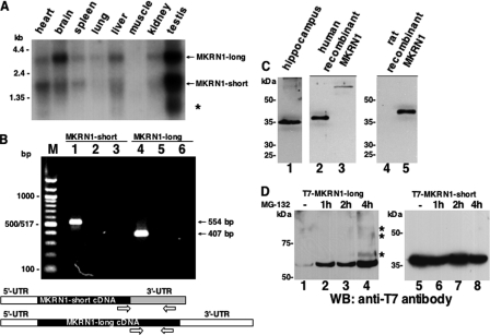
FIGURE 5.
Characterization of MKRN1 expression in rat brain at the mRNA and protein level. A, poly(A)-RNAs from various rat tissues were hybridized with a 32P-labeled MKRN1 probe that detects both MKRN1-long (3.2 kb) and MKRN1-short (2 kb) transcripts. The asterisk denotes a third transcript variant of 0.75 kb. The positions of marker RNAs (in kb) are indicated on the left. B, RT-PCR with rat hippocampal RNA was performed to confirm the expression of MKRN1-short (lane 1, 554 bp) and MKRN1-long (lane 4, 407 bp) in this brain region. The primers used for the amplification reactions (open arrows) are schematically shown in the lower part of this panel. As controls, PCRs were done in the absence of template (lanes 2 and 5) as well as with non-reverse transcribed RNA (lanes 3 and 6). PCR products were resolved by agarose gel electrophoresis along with a 100-bp ladder marker (left) followed by ethidium bromide staining. C, rat hippocampal proteins were probed with rabbit anti-MKRN1 antiserum (lane 1). Human T7-tagged MKRN1-short (lane 2) and MKRN1-long (lane 3) expressed in HEK-293 cells were run in parallel on the same gel. Antibodies raised against human MKRN1-short recognize recombinant myc-His-tagged rat MKRN1-short (lane 5). Non-transfected control extract is shown in lane 4. D, MKRN1-long is only efficiently expressed in HEK-293 cells if proteasomal degradation is inhibited by the addition of MG-132 (final concentration, 10 μm). The compound was added 1, 2, or 4 h (lanes 2–4) before protein extraction. Bands marked by asterisks most likely correspond to ubiquitinated forms of MKRN1-long. MKRN1-short lacks autoubiquitination properties, probably due to the C-terminal deletion of six amino acids of its RING finger domain. Destabilization of the short MKRN1 variant is not seen if cells are grown in the absence of MG-132 (lane 5). The addition of the compound for various periods of time does not alter the steady state concentration of MKRN1-short (lanes 6–8). The positions of molecular size marker proteins (in kDa) are indicated on the left. WB, Western blot.

