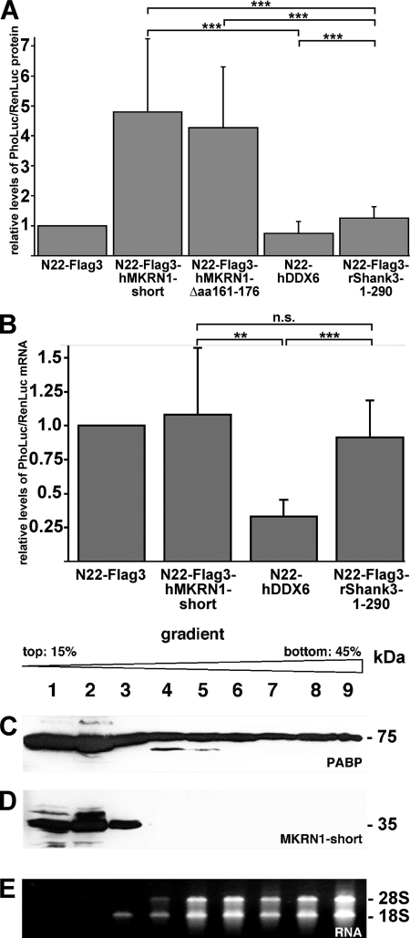
FIGURE 7.
MKRN1-short stimulates translation in nerve cells. The eukaryotic expression vector pinFiRein-boxB16B was co-transfected with vectors encoding fusion proteins consisting of an N-terminal N22 peptide and either hMKRN1-short, hMKRN1-Δ487–535, hDDX6, or rShank3–1-299 into dispersed cortical neurons at 7 DIV. The empty vector N22-FLAG3 served as a control (for details see “Experimental Procedures”). A, dual luciferase assays were performed at 9 DIV. The relative levels of PhoLuc/RenLuc proteins are shown (in arbitrary units). B, RNAs from transfected neurons were prepared on 9 DIV, transcribed into cDNAs, and subjected to real-time PCR analyses using Pho/Luc- and RenLuc-specific TaqMan assays. The relative levels of PhoLuc/RenLuc mRNAs are shown (in arbitrary units). Bars represent S.E. Statistical analyses were done using Student's t test (**, p < 0.01; ***, p < 0.001, n.s., not significant). C and D, ribosomes/polysomes from adult rat hippocampi were fractionated by sucrose gradient ultracentrifugation. Individual fractions (lanes 1–9) were subjected to SDS-PAGE and Western blot analyses using anti-PABP (C) or anti-MKRN1 antibodies (D). The positions of molecular size marker proteins (in kDa) are indicated on the right. E, RNAs purified from individual gradient fractions were separated by agarose gel electrophoresis and stained with ethidium bromide. 28 S and 18 S, large and small ribosomal RNAs.

