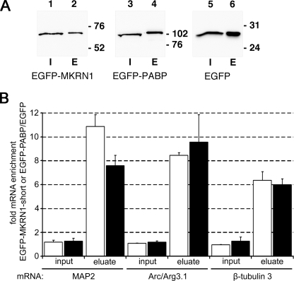
FIGURE 8.
MKRN1-short is associated with dendritically localized mRNAs. A, shown are protein lysates from rat primary cortical neurons expressing recombinant EGFP-MKRN1-short, EGFP-PABP, or EGFP alone that were subjected to immunoprecipitation with GFP Trap®_A beads followed by SDS-PAGE and Western blot analyses using anti-GFP antibodies for detection of EGFP-MKRN1 (lanes 1 and 2), EGFP-PABP (lanes 3 and 4), and EGFP (lanes 5 and 6), respectively. I, input proteins (lanes 1, 3, and 5); E, immunoprecipitated proteins eluted from the beads (lanes 2, 4, and 6). The positions of molecular size marker proteins (in kDa) are indicated on the right. B, RNAs extracted from whole cell lysates (input) and immunoprecipitated protein fractions (eluate) were subjected to semiquantitative real-time RT-PCR using primers for MAP2-, Arc/Arg3.1-, and neuron-specific β-tubulin 3 cDNAs. The graph depicts the enrichment of individual transcripts in inputs and in eluates obtained by immunoprecipitation of EGFP-MKRN1-short (open bars) and EGFP-PABP (closed bars), respectively, compared with the empty vector control (EGFP-IP). Data were obtained using REST (relative expression software tool) 2008 software for group-wise comparison and statistical analysis of relative expression results in real-time PCR (41). S.E., vertical lines, enrichment/eluates, p < 0.001; enrichment/inputs, not significant. Arc/Arg3.1, activity-regulated cytoskeleton-associated.

