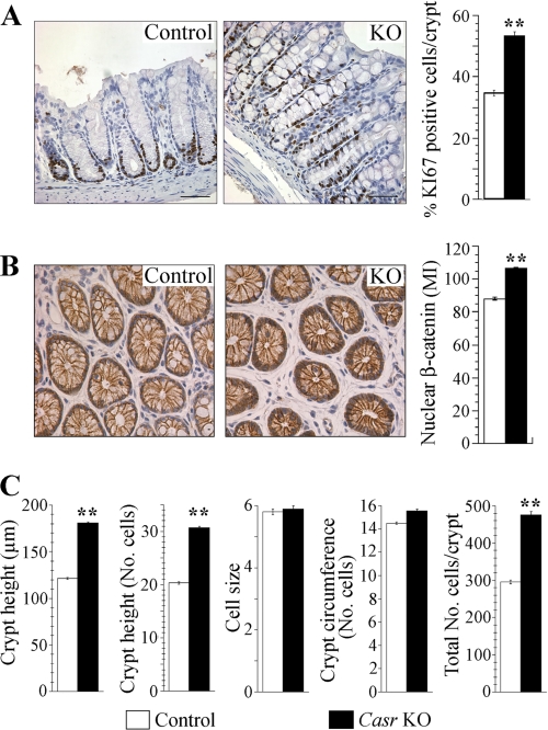
FIGURE 2.
Proliferation analysis of colon sections of Casr KO mice. A, colons fixed and processed for immunocytochemistry using a Ki-67 antibody as described under “Experimental Procedures.” Ki-67-positive cells were quantified in a sample size of 5 mice/group with at least 20 crypts examined per mouse. Data from KO and control littermates were represented as mean of the percentage of Ki-67-positive cells ± S.E. and compared by unpaired Student's t test (**, p < 0.0001). Scale bars, 10 μm. B, quantification of nuclear β-catenin in colonocytes of Casr KO mice. The same level cross-sections of colonic crypts were processed for immunocytochemistry using a β-catenin rabbit antibody as described under “Experimental Procedures.” Quantification was performed as described under “Experimental Procedures” in a sample size of 3 mice/group with at least 60 cells examined per animal. Values represent the MI of nuclear β-catenin ± S.E., and they were compared using unpaired Student's t test (**, p < 0.0001). C, morphometric analysis of Casr KO colonic crypts. Full-length, longitudinally cut crypts (at least 20 per mouse) from 5 Casr KO and 5 control littermates were analyzed for crypt height (micrometers) and number of cells per crypt height. Cross-section of crypts (20/mouse) was used to determine the average crypt diameter (micrometers) and circumference (in number of cells). These data were used to calculate cell size (crypt height in micrometers/crypt height in cell number) and estimate the total cells per crypt (mean cells per crypt column × mean crypt circumference). Data from KO and control littermates are represented as mean ± S.E. and compared using unpaired Student's t test (**, p < 0.0001).

