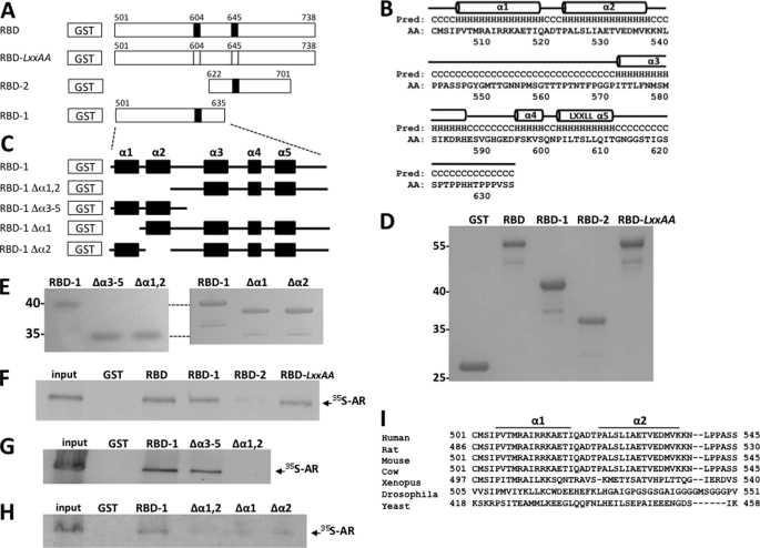
FIGURE 5.
AR binds to a novel noncanonical α-helical array in the MED1 RBD. A, schematic representation of the GST-MED1-RBD (amino acids 501–738) and mutant derivative fusion proteins. LXXLL motifs are indicated by black bars; LXXLL to LXXAA are indicated by open bars. B, predicted secondary structure of MED1 amino acid residues 501–635. Five α-helical motifs (α1, α2, α3, α4, and α5) are indicated by horizontal cylinders. C, schematic representation of the GST-MED1-RBD1 (amino acids 501–635) and mutant derivative fusion proteins. The α-helices α1, α2, α3, α4, and α5 are indicated by black boxes. D and E, purified GST-MED1 fusion proteins were fractionated by SDS-PAGE and stained with Coomassie blue. F–H, GST pulldown assays were carried out by incubating [35S]methionine-labeled AR (labeled in the presence of 10 nm DHT) together with GST-MED1-RBD and mutant derivative fusion proteins (see A and C). The bound proteins were detected by autoradiography. I, sequence alignment of the identified AR-binding noncanonical α-helical array in MED1 of different species.

