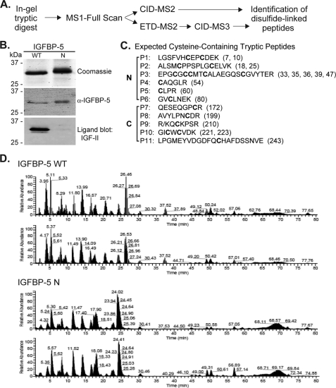
FIGURE 1.
Experimental plan for mapping disulfide linkages of IGFBP-5. A, analytical approach for identification of disulfide linkages in IGFBP-5 by MS. See “Experimental Procedures” for details. B, analysis of purified wild type and N-terminal mutant IGFBP-5 after non-reducing SDS-PAGE by staining with Coomassie Blue (top), immunoblotting with anti-IGFBP-5 antibody (middle), and ligand blotting with biotinylated human IGF-II (bottom). C, list of the 11-tryptic peptides (P1-P11) in mouse IGFBP-5 that contain cysteine residues (cysteines are in bold script) along with their location in the N- or C-terminal domain of the protein. Numbers in parentheses denote the location of each cysteine in the amino acid sequence. D, analysis of IGFBP-5 tryptic peptides by MS. Elution profiles after separation by liquid chromatography for 0 to 80 min of peptides/ions derived from trypsin digestion of wild type (top) or N-mutant IGFBP-5 (bottom). Results of two independent experiments for each IGFBP-5 species are shown and demonstrate the reproducibility of each protein's tryptic peptide profile.

