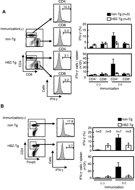
Figure 3.
IFN-γ production by CD4 splenocytes from LM secondarily infected HBZ-Tg mice decreases in CD4+ Foxp3− T cells. Mice were immunized and challenged as shown at the top of Figure 2B, and their splenocytes were harvested at 12 hours after challenge and analyzed for intracellular IFN-γ production. (A) Splenocytes were gated by CD3 expression, and IFN-γ production was measured in living CD4 or CD8 T cells using FACS. (B) IFN-γ production in CD3+ CD4+ Foxp3− cells was determined. Bars represent the mean ± SD of all mice per genotype. Two independent experiments have been performed.

