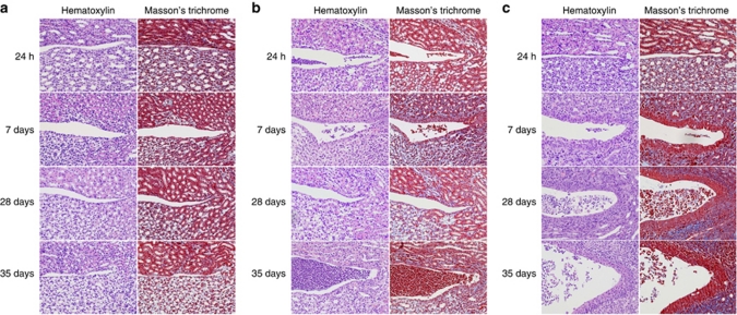
Figure 5.
Epithelial hypertrophy and tissue fibrosis in mCxcr2+/+, mCxcr2+/−, and mCxcr2−/− mice. (a) Minor epithelial proliferation in mCxcr2+/+ mice, 7 days after infection, but no evidence of fibrosis. (b) In mCxcr2+/− mice, subepithelial fibrosis and epithelial hypertrophy was observed 7 days after infection. Proliferating epithelium was seen after 14 days of infection in the mCxcr2+/− mice. (c) Thickening of the pelvic epithelium and fibrosis were observed 7 days post infection in the mCxcr2−/− mice. After 28 days, the inflammatory cell infiltrate was abundant and Russell bodies were observed. At 35 days post infection, the tissue was destroyed and replaced by abscesses and inflammatory cells. Proliferation is visualized by hematoxylin–eosin staining, and Masson's trichrome was used to visualize tissue fibrosis (blue).

