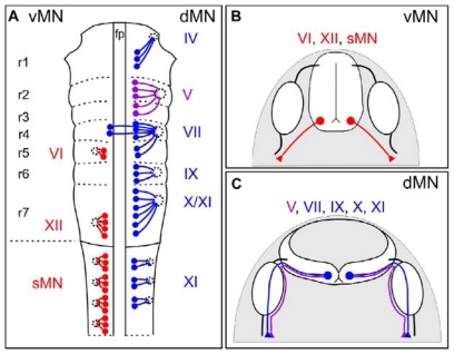
Figure 1.
Motor neuron subtypes and the projections of their axons out of the CNS. (A) Schematic of motor neuron nuclei in the developing brainstem (rhombomere (r), r1 to r7) and spinal cord. vMNs are indicated in red on the left, whereas dMNs are indicated in blue on the right of the schematic. Trigeminal (V) motor nuclei are shown in purple. Each cranial motor nuclei is numbered in roman numerals, e.g., CN XI. Abbreviations: fp, floor plate; sMN, spinal motor neuron; (B) Axonal projections of vMNs in the hindbrain (VI, XII) and spinal cord (sMN) are shown in red; (C) Axonal projections of dMNs (VII, IX, X, XI) and trigeminal (V) dMNs are shown in blue and purple, respectively. Note that axons extending from trigeminal dMNs avoid sensory ganglia (white ovals), while axons of other dMN invade these ganglia [6].

