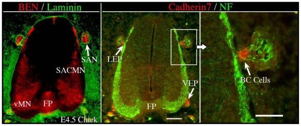
Figure 2.
Cadherin 7-expressing BC cells are located at both the lateral and ventral exit points in chick embryos. Cervical spinal cord-containing transverse cryosections derived from E4.5 chick embryos were doubled-labeled with either anti-Ben and anti-Laminin (Left) or anti-Cadherin 7 and anti-NF (Middle, Right), and the appropriate secondary antibodies. (Left) BEN is expressed by both SACMN and vMN at this developmental stage, the FP and the SAN, which is positioned outside and adjacent to the spinal cord. Anti-Laminin labeling demarcates the margin of the spinal cord. (Middle) Cadherin 7-expressing BC cells are located at both the LEP and VEP. In this panel, the SAN is labeled by anti-NF. (Right) A higher magnification view of the boxed area in the middle panel. SACMN, spinal accessory motor neurons; SAN, spinal accessory nerve; vMN, ventral motor neurons; FP, floor plate; LEP, lateral exit point; VEP, ventral exit point; BC cells, boundary cap cells. Scale bar in middle panel, 100μm, applies to the left and middle panels. Scale bar in right panel, 50 μm.

