
Figure 3.
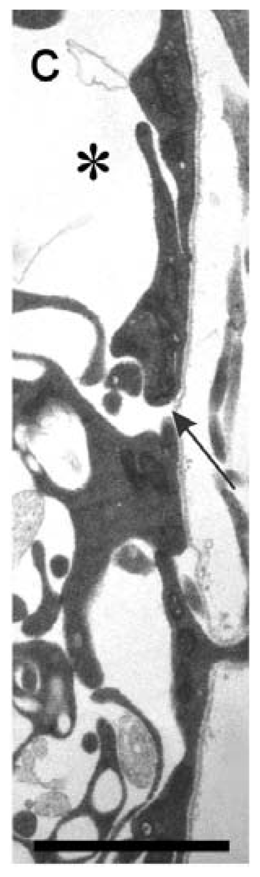
Electron micrograph of a section of neural tube derived from an E11 rat embryo at a presumptive motor exit point. A transverse section of the neural tube shows a thin layer of uninterrupted basal lamina that overlies glial end-feet, which form a conspicuous gap at a presumptive motor exit point. Black arrow, presumptive motor exit point; black asterisk, space found within presumptive white matter. Scale bar: 2 μm [42].